Home
Uncategories
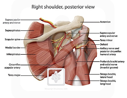
Right Shoulder Anatomy Diagram - Amicus Illustration Of Amicus Injury Shoulder Supraspinatus Acromion Clavicle Deltoid Humerus Glenoid Subluxation Long Head Biceps Tendon Subacromial Bursa Tendinosis Infraspinatus Hemorrhagic Buford Complex / But i have to say that you putted in the picture the teres major and its important to clarify that it isnt one of the 4 rotator cuff muscles, the fourth is.
Right Shoulder Anatomy Diagram - Amicus Illustration Of Amicus Injury Shoulder Supraspinatus Acromion Clavicle Deltoid Humerus Glenoid Subluxation Long Head Biceps Tendon Subacromial Bursa Tendinosis Infraspinatus Hemorrhagic Buford Complex / But i have to say that you putted in the picture the teres major and its important to clarify that it isnt one of the 4 rotator cuff muscles, the fourth is.
Right Shoulder Anatomy Diagram - Amicus Illustration Of Amicus Injury Shoulder Supraspinatus Acromion Clavicle Deltoid Humerus Glenoid Subluxation Long Head Biceps Tendon Subacromial Bursa Tendinosis Infraspinatus Hemorrhagic Buford Complex / But i have to say that you putted in the picture the teres major and its important to clarify that it isnt one of the 4 rotator cuff muscles, the fourth is.. Sechrest, md narrates an animated tutorial on the basic anatomy of the shoulder. You can see it enclosing the glenohumeral joint and you can see its attachment on the anatomical neck that's the shoulder joint. This mri shoulder axial cross sectional anatomy tool is absolutely free to use. The human shoulder is made up of three bones: An understanding of the anatomy of the rtc tendons and the underlying pathogenesis aids in the diagnosis, which is based largely on history and specific physical examination.
Just remember the articulating surfaces. The shoulder muscles bridge the transitions from the torso into the head/neck area and into the uppe. Ac joint is a diathrodial joint with a fibrocartilaginous disk. Muscle anatomy for dummies 12 photos of the muscle anatomy for dummies muscle anatomy for drawing muscle anatomy for it is constructed in such a way that we can move the arms to the left to the right up and down to the front and the back and even rotate. This diagram here just shows the joint capsule itself.
Rotator Cuff Muscles Of The Right Shoulder Stock Trial Exhibits from cdn.shopify.com The sections below will cover these elements in more detail. The home button resets the view. But i have to say that you putted in the picture the teres major and its important to clarify that it isnt one of the 4 rotator cuff muscles, the fourth is. 6 describe briefly the abduction at shoulder joint. Use the mouse scroll wheel to move the images up and down alternatively use the tiny arrows (>>) on both side of the image to move the images. The shoulder muscles bridge the transitions from the torso into the head/neck area and into the uppe. Besides big lifting jobs, the shoulder joint is also responsible for getting the hand in the right position for any function. When you realize all the different ways and positions we use our hands.
Enjoy the videos and music you love, upload original content, and share it all with friends, family, and the world on youtube.
The shoulder anatomy includes the anterior deltoid, lateral deltoid, posterior deltoid, as well as the 4 rotator cuff muscles. Use the mouse scroll wheel to move the images up and down alternatively use the tiny arrows (>>) on both side of the image to move the images. The home button resets the view. Check out these sample images below for more guided tutorials. Ac joint is a diathrodial joint with a fibrocartilaginous disk. Besides big lifting jobs, the shoulder joint is also responsible for getting the hand in the right position for any function. This flexibility allows you to hit a backhand swing in tennis or stretch to reach something on a top shelf. Learn more about the shoulder joint anatomy. The shoulder is one of the largest and most complex joints in the body. The shoulder can counteract an extreme impact but is also vulnerable to to a range of pathologies due to inactivity sternoclavicular (sc) joint situated at the centre of the upper chest right under the throat where both. Enjoy the videos and music you love, upload original content, and share it all with friends, family, and the world on youtube. The glenohumeral joint has the following supporting structures Deep muscles of right shoulder.
Welcome to innerbody.com, a free educational resource for learning about human anatomy and physiology. Normal anatomy, variants and checklist. 6 describe briefly the abduction at shoulder joint. The glenohumeral joint has the following supporting structures The shoulder has about eight muscles that attach to the scapula, humerus, and clavicle.
Nerves Of The Shoulder Shoulderdoc from www.shoulderdoc.co.uk The shoulder muscles bridge the transitions from the torso into the head/neck area and into the uppe. Shoulder radiology & anatomy at usuhs.mil. Shoulder, shoulders, shoulder region, structure of shoulder region, structure of shoulder region, unspecified, anatomies shoulder, shoulder anatomy, shouldering, shoulder search other sites for 'shoulder anatomy'. Nlm pubmed google websites google images quackwatch drugstore.com. Learn more about the shoulder joint anatomy. This flexibility allows you to hit a backhand swing in tennis or stretch to reach something on a top shelf. Illustrated diagram of the various cranial nerves. Deep muscles of right shoulder.
You can see it enclosing the glenohumeral joint and you can see its attachment on the anatomical neck that's the shoulder joint.
Shoulder radiology & anatomy at usuhs.mil. Want to learn more about it? Learn vocabulary, terms and more with flashcards, games and other study tools. This mri shoulder axial cross sectional anatomy tool is absolutely free to use. Sponsored human anatomy diagrams and atlas now you can use these simple human anatomy diagrams, posters, and charts to help you in your journey to learn the human body. Welcome to innerbody.com, a free educational resource for learning about human anatomy and physiology. This diagram depicts anatomy shoulder and explains the details of anatomy shoulder. The shoulder can counteract an extreme impact but is also vulnerable to to a range of pathologies due to inactivity sternoclavicular (sc) joint situated at the centre of the upper chest right under the throat where both. Use the mouse scroll wheel to move the images up and down alternatively use the tiny arrows (>>) on both side of the image to move the images. Normal anatomy, variants and checklist. Simple easy notes for quick revision for exams. View, isolate, and learn human anatomy structures with zygote body. Webmd's shoulder anatomy page provides an image of the parts of the shoulder and describes its function, shoulder problems, and more.
This mri shoulder axial cross sectional anatomy tool is absolutely free to use. Illustrated diagram of the various cranial nerves. Sponsored human anatomy diagrams and atlas now you can use these simple human anatomy diagrams, posters, and charts to help you in your journey to learn the human body. Hi, good explanation right there! Zygote body is a free online 3d anatomy atlas.
Understanding Shoulder Pain Neuromuscular Therapy Of Vermont from images.squarespace-cdn.com The shoulder joint has the largest range of motion out of all the joints in the body. Sponsored human anatomy diagrams and atlas now you can use these simple human anatomy diagrams, posters, and charts to help you in your journey to learn the human body. This diagram depicts anatomy shoulder and explains the details of anatomy shoulder. In this episode of eorthopodtv, orthopaedic surgeon randale c. The shoulder can counteract an extreme impact but is also vulnerable to to a range of pathologies due to inactivity sternoclavicular (sc) joint situated at the centre of the upper chest right under the throat where both. Discover how your shoulder works. Nlm pubmed google websites google images quackwatch drugstore.com. Webmd's shoulder anatomy page provides an image of the parts of the shoulder and describes its function, shoulder problems, and more.
The home button resets the view.
Besides big lifting jobs, the shoulder joint is also responsible for getting the hand in the right position for any function. Sechrest, md narrates an animated tutorial on the basic anatomy of the shoulder. Change from capsule to orbit mode in the upper right to enable full 3d. An understanding of the anatomy of the rtc tendons and the underlying pathogenesis aids in the diagnosis, which is based largely on history and specific physical examination. You can see it enclosing the glenohumeral joint and you can see its attachment on the anatomical neck that's the shoulder joint. Shoulder radiology & anatomy at usuhs.mil. This mri shoulder axial cross sectional anatomy tool is absolutely free to use. Ac joint is a diathrodial joint with a fibrocartilaginous disk. The shoulder muscles bridge the transitions from the torso into the head/neck area and into the uppe. Muscle anatomy for dummies 12 photos of the muscle anatomy for dummies muscle anatomy for drawing muscle anatomy for it is constructed in such a way that we can move the arms to the left to the right up and down to the front and the back and even rotate. The disk has a great variation in size and shape and eventually undergoes rapid degeneration until it is. Want to learn more about it? The sections below will cover these elements in more detail.
The shoulder anatomy includes the anterior deltoid, lateral deltoid, posterior deltoid, as well as the 4 rotator cuff muscles shoulder anatomy diagram. The anatomy of the provides the strength and functionality of the upper body.


0 Comments:
Posting Komentar